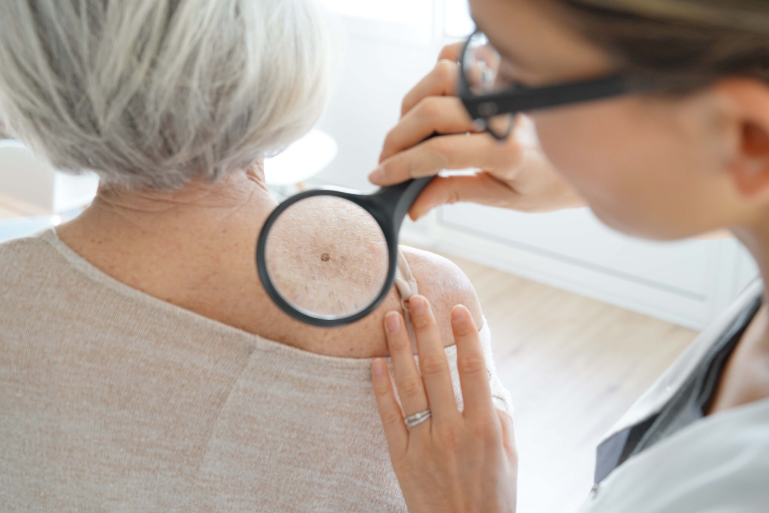
Mohs surgery represents a precise treatment method for removing skin cancer while preserving healthy tissue. This specialized surgical technique removes skin cancer layer by layer, examining each layer under a microscope until all cancer cells are eliminated. This approach offers a solution for various types of skin cancer, particularly basal cell carcinoma and squamous cell carcinoma. Here are the general steps of this surgery:
1. Cleansing & Numbing
The surgical team begins by thoroughly cleaning the treatment area, typically with an antiseptic solution. Local anesthesia is administered to completely numb the skin and surrounding tissue. This preparation phase is designed to make the procedure more comfortable for you, allowing the surgeon to work with precision.
The numbing medication takes effect before the surgery. The surgeon marks the visible tumor boundaries with directional references to guide the surgical area. While the anesthesia works to eliminate sensation, the medical team prepares the necessary surgical instruments.
2. Taking Tissue Sample
After the area becomes completely numb, the surgeon may remove the visible tumor along with a thin layer of surrounding healthy tissue. The tissue sample is carefully mapped and marked to track its exact location on your body. This mapping process allows for precise reconstruction later.
The removed tissue goes directly to the on-site laboratory, and the wound is temporarily bandaged. The surgeon may take detailed notes about the specimen’s orientation and location to maintain accuracy throughout the process. This immediate analysis helps guide the surgeon in making decisions during the procedure to make sure all cancerous tissue has been removed.
3. Freezing the Tissue
Laboratory technicians typically rapidly freeze the tissue sample and prepare it for microscopic examination. During Mohs surgery, the frozen tissue is sliced into thin sections, revealing cellular details clearly. While you wait, a specialist typically stains these tissue sections with special dyes that highlight cancer cells. This freezing process preserves the tissue’s cellular structure. The laboratory staff works to prepare slides that show whether cancer cells remain at the tissue margins.
4. Checking & Repeating Process
The Mohs surgeon examines each tissue section under a high-powered microscope. When cancer cells appear at the margins of the removed tissue, the surgeon marks these areas on the detailed map created earlier. The surgeon returns to remove additional tissue only from the marked areas, allowing for a targeted approach that preserves healthy skin.
If the microscopic examination reveals clear margins without cancer cells, the procedure is complete. The surgeon discusses reconstruction options with you, which may include allowing the wound to heal naturally, closing it with stitches, or performing a skin graft, depending on the wound’s size and location.
- Quick results: The entire process typically completes within six hours
- Precise removal: Only cancerous tissue is removed, preserving healthy skin
Schedule Mohs Surgery Today
Mohs surgery offers an effective treatment option for many types of skin cancer. The procedure’s systematic approach removes cancer while minimizing damage to surrounding healthy tissue. Your dermatologist will determine if Mohs surgery suits your specific situation based on factors like cancer type, location, and size. Contact your healthcare provider to discuss whether Mohs surgery is a suitable treatment option for your skin cancer. Early treatment often leads to better outcomes, so don’t delay in seeking professional medical advice about your skin health concerns.


